Postagens

The plant cell

Imagem
1- Nucléolo ( Nucleolus) 2- Núcleo (Core) 3- Cloroplasto (Chroloroplast) 4- Vacúolo (Vacuole) 5- Complexo de Golgi (Golgi Complex) 6- Amiloplastos (Amyloplasts) 7- Mitocôndria (Mitochondria) 8- Membrana Plasmática (Plasma Membrane) 9- Lisossomo (Lysosome) 10- Retículo Endoplasmático (Endoplasmatic Reticulum)  11- Citoplasma (Cytoplasm) 12- Ribossomo (Ribosome) The plant cell has the presence of the outer cellulose cell wall that gives the defined shape. In addition, it has a large vacuole that contains water and dissolved substances, another feature is the presence of plastids, among them the chloroplasts responsible for photosynthesis. Colorless plasters and leucoplasts can also be found with the storage function.   References: SCHWAMBACH, C; SOBRINHO, G. C. Animal and Plant Research. 1st edition. Editora Érica, 2015. 192 p.

Ribosomes

Imagem
Ribosomes are membraneless granules composed of RNA molecules associated with proteins, present in all cells. They are responsible for the synthesis of the proteins, by a single group of amino acids, by a control controlled by the mRNA formed in the genetic transcription, by a command of the DNA. Reference: AMABIS, José Mariano; MARTHO, Gilberto Rodrigues. Biologia das Células 1. 4th edition. São Paulo: Editora Moderna, 2015. v.1, 528 p.

Cytoskeleton

Imagem
Cytoskeleton is a complex network of proeins that determines the shape of cells. Ii is fundamental for cellular moviment to it  occur, provides the structural support and mobility of intracellular organelles and the structure by moviment and separtion of chromosomes during cell division. The main components os the cytoskeleton are: mecrofilaments, filaments and microtubules.    Reference:MONTEIRO. M.R , Artigos Científicos , Journal of Empilepsy and Clinical Neurophysiology 2011  PAG -122 

Gastrulation

Imagem
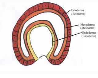
Gastrulation is the process that orginates the gern layers ( ectoderm,  mesoderm and endoderm.) While this process happens the embrionic disc, enitially bilaminar, is coverted into a trilaminar embrionic disc. During gastrulation the cells move into the embryo and the regions of the endoderm and mesoderm which are originally adjacent to the cell plate, they assume theirs apropiate positions in the ambryo.  Reference-WOLPERT.  L . Princípios de Biologia do desenvolvimento . Porto alegre , 2000. Pag- 484  

The Plasma Membrane

Imagem
The plasma membrane maintains the medium has receptors fot hormones and other chemical signals establishing connections of the cells with each other . Its outer surface is richn protein and lipid molecules contaning glucose. It is responsible for fotming adhesion structures of intracellular space and communicetion btween cells . It is the main responsible for the control of penetration and exit of substances from the cell.  It consists of a medium hydrophilic layers; yhe peripheral proteins areon the cytoplasmic face of the membrane, and the same trailaminar structure is seenin all cell membranes. Reference-REZEK,Angelo Junqueira. Biologia Celular e Molecular 9° edição Guanabara Koogan. Pag 81-105 , 01/2012 .

Embryonic leaflets

Imagem
 An embryonic leaflet is the primary layer of cells that will subsequently give rise to all other tissues and organs of animals during the process of organogenesis. The three embryonic leaflets, or germ layers, form during embryogenesis and, in vertebrates, are particularly pronounced. However, all Eumetazoas  produce two or three embryonic leaflets. Animals with radial symmetry, such as Cnidarians, produce two layers (the ectoderm and the endoderm) leading to the classification of diblásticos. Animals with bilateral symmetry produce a third camanda (the mesoderm), between the two other layers, leading to the classification of triblásticos.    They are formed in the second week of development of the embryo in the process of Gastrulation, where the formation of germ layers will occur.   Ectoderm will give rise to:   Epidermis and cutaneous attachments, all structures of the nervous system (brain, nerves, nerve ganglia and spinal cord) and epithel...

Mitochondria

Imagem
1.         Granules 2.         Crests 3.         Particle  synthesizers of  atp 4.         Complex  fs 5.         Intermembranous  space 6.         Mitochondrial  dna 7.         Internal  mitochondrial  membrane 8.         Outer  mitochondrial  membrane 9.         Ribosomes 10.      Matrix   Mitochondria is a membranous cytoplasmic organelle whose main function is the generation of energy through the synthesis of adenosine triphosphate   Inside the mitochondria occurs cellular respiration, a process in which organic molecules of...